Lv Size Echo


Aug 27, 2009 · Echo studies have generally lv size echo defined thrombus as a mass within the LV cavity with margins distinct from ventricular endocardium and distinguishable from papillary muscles, chordae, trabeculations, or technical artifacts. 5,6 Additional features, such as pattern of mobility, 7 size, 8 and associated ventricular wall motion abnormalities, 9–13 ...
Echo assessment of LV size and mass and their changes over time predict risk of ventricular arrhythmias post MI; postinfarction remodeling may be an important substrate for triggering such arrhythmias
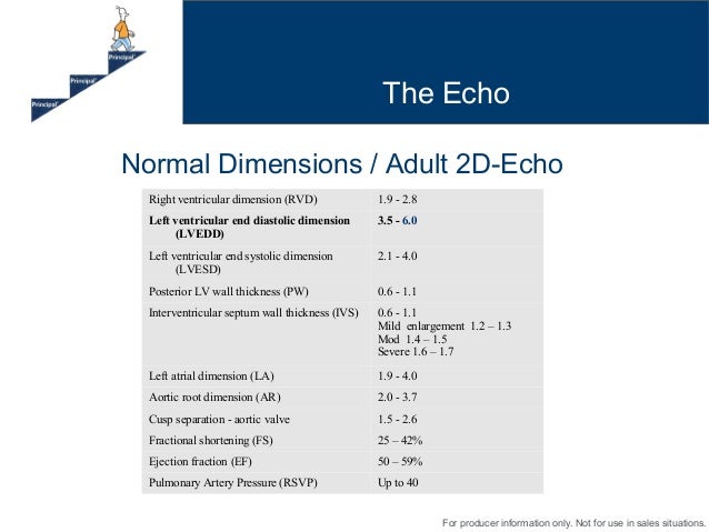
Measuring left ventricular size and wall thickness is a standard part of the routine echo examination. There are normative values for LV wall thickness, and the trainee sonographer is taught basic pattern-recognition in the early phases of training to identify patients with left ventricular hypertrophy.
Size left ventricle. ... In European journal of echocardiography : the journal of the Working Group on Echocardiography of the European Society of Cardiology 11 (3), pp. 223–244. DOI: 10.1093/ejechocard/jeq030. –>Pubmed-Link. Pulmonary valve stenosis …
Dec 01, 2016 · Methods. This study was performed on 1,364 healthy adults aged 18–35 years. Standard trans-thoracic echocardiographic studies were performed to obtain end diastole measurements of left ventricular (LV) posterior wall thickness (PWd), interventricular septum thickness (IVSd), LV internal dimensions at end diastole (LVEDD) and end systole (LVESD), left atrial (LA) diameter, aortic root ...
Ethnic-Specific Normative Reference Values for ...
Ethnic-Specific Normative Reference Values for Echocardiographic LA and LV Size, LV Mass, and Systolic Function: The EchoNoRMAL Study JACC Cardiovasc Imaging . 2015 Jun;8(6):656-65. doi: 10.1016/j.jcmg.2015.02.014.LV End Diastolic Diameter cm - E-Echocardiography
LV Volume = [7/(2.4 + LVID)] * LVID 3. RWMA, either close or distant, may cause the volume analysis to be incorrect. If the endocardial boarder is poorly seen, then the area of the left ventricular cavity may be inaccurate. Failing hearts (i.e. cardiomyopathy) tend to be more spherical than normal hearts so the spherical formula may be more ...Understanding LVH Part 2: How to Measure LV Mass and ...
LVM is the acronym lv size echo for Left Ventricular Mass. LV mass (LVM) is a vital prognostic measurement we obtain with echocardiography to manage hypertension. RWT is the acronym for Relative Wall Thickness and is an additional reference value that can help further classify the type of LVH.Over 150,000 platelets were individually tracked in each LV model over 15 cardiac cycles. As LV size decreased, platelets experienced markedly lv size echo increased shear stress histories (SHs), whereas platelet residence time (RT) in the LV increased with size.
RECENT POSTS:
- louis garneau tri clothing
- cheap cardinals tickets az
- authentic louis vuitton pochette metis
- louis vuitton hat box pictures
- louis vuitton toiletry pouch 15 vs mini pochette
- is louis vuitton bags ever made in the usa
- small canvas pouch bags
- louis vuitton blue monogram sweater
- laser metal engraving machine cheap
- cheap hotel rooms downtown st louis
- ebay louis vuitton mens bag
- louis vuitton bags paris
- authentic hermes birkin bag
- singapore calendar 2020 with public holidays and school holidays
All in all, I'm obsessed with my new bag. My Neverfull GM came in looking pristine (even better than the Fashionphile description) and I use it - no joke - weekly.
Other handbag blog posts I've written:
louis vuitton monogram canvas boulogne 35 bag
louis vuitton monogram logomania grey scarf
Do you have the Neverfull GM? Do you shop pre-loved? Share your tips and tricks in the comments below!
*Blondes & Bagels uses affiliate links. Please read the replica lv wallets for women for more info.